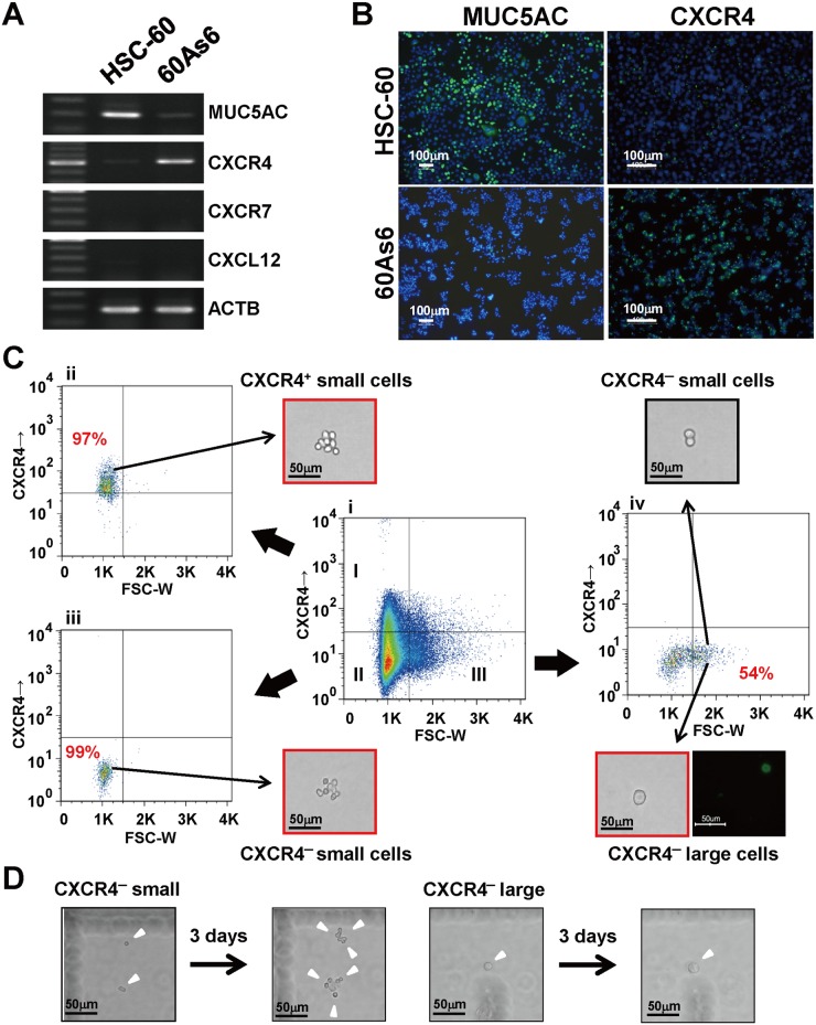
Fig 1. Expression of CXCR4 is upregulated after serial transplantation.
(A) RT-PCR of MUC5AC, CXCR4, CXCR7, and CXCL12 in a GC cell line HSC-60 and its subline 60As6. (B) Immunocytochemistry for MUC5AC (left panel, green) and CXCR4 (right panel, green) in HSC-60 and 60As6 cells, respectively. Nuclei were counterstained with DAPI (blue). Scale bars represent 100μm. (C) Flow cytometry analysis of cell-surface CXCR4 in 60As6 cells. The inner panels represent CXCR4+ small cells (I), CXCR4- small cells (II) and CXCR4- large cells (III) (Ci). Each cell population was separated by FACS at its respective purity (Ci-Civ). Immunopositive cells for MUC5AC were observed in only the CXCR4- large cell fraction (Civ, lower panel, green). Scale bars represent 50μm. (D) Representative images after culturing CXCR4- small and large cells for 3 days. Scale bars represent 50μm.

