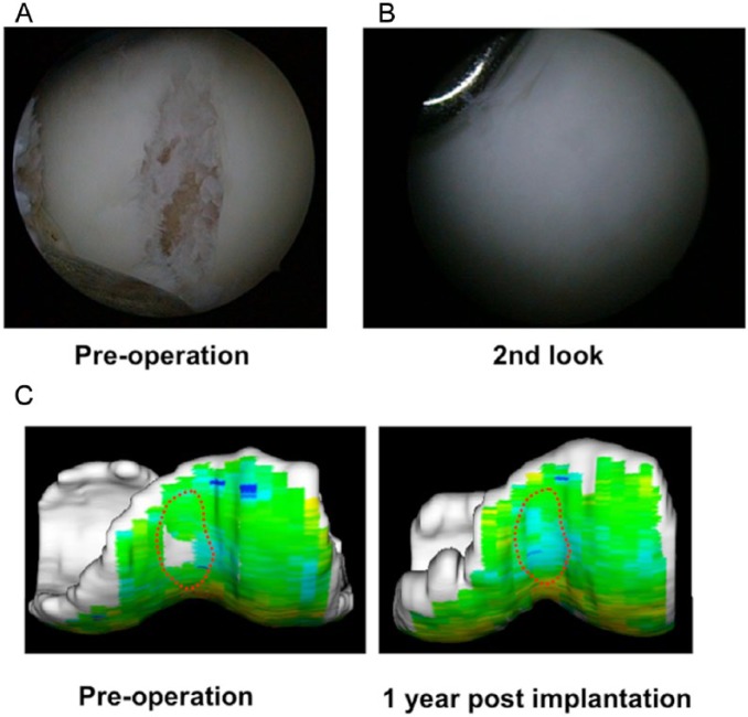
Figure 10.
Arthroscopic and magnetic resonance imaging (MRI) analyses of repair tissue following implantation of a tissue-engineered construct (TEC) to repair human chondral defects in clinical trial. (A, B) Arthroscopic views of the preoperation defect and then 1 year after implantation of a TEC. The cartilage defect was completely covered with a cartilage-like repair tissue. (C) T2-weighted mapping of the lesion at the femoral groove. Left, before implantation; right, 1 year after implantation.

