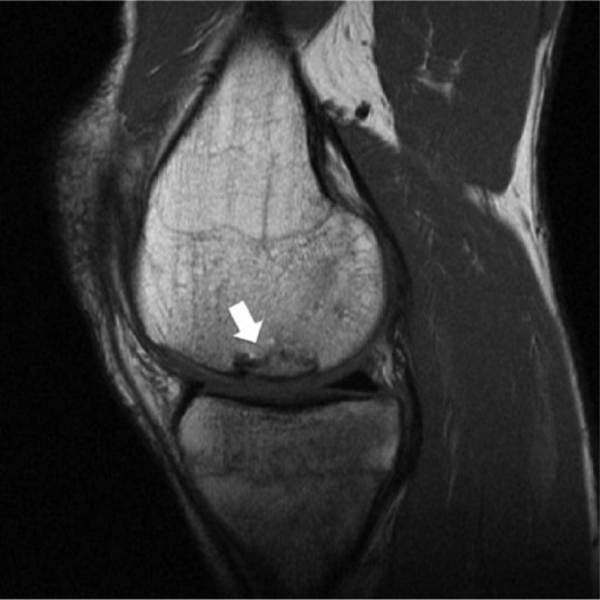
Figure 2.

Sagittal intermediate-weighted magnetic resonance image of a patient after osteochondral allograft (OCA) transplantation in the medial femoral condyle (arrow). OCA cartilage signal, “fill,” and surface congruity are normal compared with adjacent host cartilage. Subchondral bone plate is incongruent, but subchondral bone marrow signal is preserved and the host-graft junction demonstrates some osseous incorporation.
