
Figure 1.
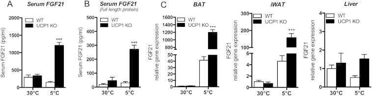
Cold induced increase of FGF21 serum levels and gene expression in UCP1 KO mice compared to WT littermates. Mice were maintained at 30 °C or exposed to 5 °C for 3 weeks (upon acclimation to 18 °C for 2 weeks). (A) Serum levels of FGF21 and (B) active FGF21 of WT or UCP1 KO mice. (C) quantitative PCR (qPCR) analysis of FGF21 in brown adipose tissue (BAT), inguinal white adipose tissue (iWAT) and liver. ***P < 0.001, significant differences between the genotypes. Data are means ± SEM (n = 6–8/group).