Figure 7.

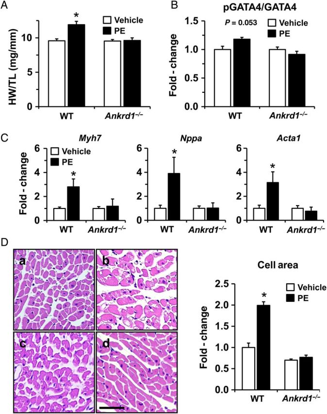
PE-induced cardiac hypertrophy is abrogated in Ankrd1−/− mice. (A) PE infusion for 2 weeks resulted in a significant increase in the ratio of heart weight to tibia length (HW/TL) when compared with vehicle controls in WT but not Ankrd1−/− mice (n = 6 in WT groups, n = 7 in Anrkd1−/− groups). (B) Ankrd1−/− mice exhibit a blunted response to PE-induced GATA4 phosphorylation, n = 3. (C) The PE-induced increase in mRNA levels for fetal gene markers Myh7, Nppa, Acta1 was attenuated in Ankrd1−/− mice, n = 5. Values expressed as mean ± SEM, *P < 0.05 vs. vehicle controls. (D) Histological sections stained with haematoxylin and eosin (H & E) are shown at ×40 magnification; a = WT vehicle, b = WT PE, c = Ankrd1−/− vehicle, d = Ankrd1−/− PE. Scale bar = 20 µm. Cardiomyocyte cross-sectional area was quantified and fold-change values expressed as mean ± SEM, n = 3, *P < 0.05 vs. vehicle control.
