
Fig. 5.
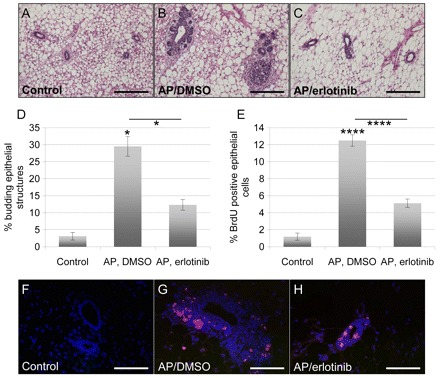
Erlotinib inhibits FGFR1-induced mammary epithelial cell budding and proliferation in vivo. (A–C) Representative images of H&E-stained sections of paraffin-embedded mammary glands from wild-type (control) mice (A), and MMTV-iFGFR1 transgenic mice treated with 1 mg per kg of body weight per day AP through intraperitoneal injections and either DMSO (B) or 25 mg per kg of body weight per day erlotinib (C) through oral gavage. Scale bars: 50 μm. (D) Quantification of the number of budding ductal structures. Every ductal structure distal to the lymph node was counted in H&E-stained sections of the 3 classes of mice, and was then grouped into budding or not budding structures. At least three sections were analyzed per mouse. Error bars represent s.e.m. *P<0.05. (E) Cellular proliferation was measured by immunofluorescence through BrdU staining. All epithelial cells were counted in ten ductal structures per section through DAPI staining, and the percentage of BrdU-positive epithelial cells was determined. A minimum of 1500 cells was counted per treatment group. Error bars represent s.e.m. ****P<0.001. (F–H) Representative images of BrdU-stained (red) sections of paraffin-embedded mammary glands from wild-type (control) mice (F), and MMTV-iFGFR1 transgenic mice treated with 1 mg per kg of body weight per day AP through intraperitoneal injections and either DMSO (G) or 25 mg per kg of body weight per day erlotinib (H) through oral gavage. The sections were also stained with DAPI (blue) to visualize all cells. Scale bars: 50 μm.