
Fig. 1. Prevailing models of symmetry breakage in the frog Xenopus.
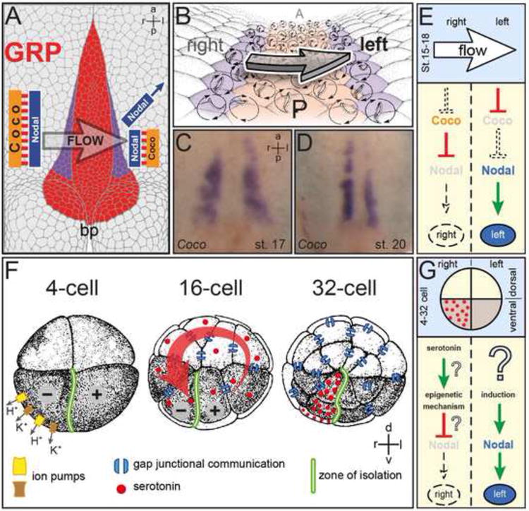
(A-E). Leftward flow. (A) Schematic representation of a stage 17 archenteron roof in ventral perspective. Flow occurs from the right to the left side of the ciliated gastrocoel roof plate (GRP; red). Nodal and Coco are co-expressed at the lateral GRP margins on both sides (purple). Flow represses Coco, activating Nodal by release of repression. bp, blastopore. (B) GRP at higher magnification. Polarized and flow-producing cilia at the GRP center are bordered by Nodal/Coco-positive cells (purple) which harbor unpolarized, sensory cilia. (C, D) Coco expression during (C) and following (D) leftward flow. Note the decrease in signal intensity on the left at post-flow stage 20 (D). (E) Schematic depiction of events on the left and right side leading up to asymmetric Nodal cascade induction in the left lateral plate mesoderm (LPM).
(F, G). Ion-flux. (F) Asymmetrically expressed ion pumps create a voltage gradient in the 4-cell embryo which initiates the electrogenic transfer of serotonin through gap junctional communication to the ventral-right lineage at the 32-cell stage. Serotonin accumulates in this lineage because the ventral midline is devoid of GJC. (F) Schematic depiction of events on the left and right side leading up to asymmetric Nodal cascade induction in the left LPM. Question marks indicate unproven interactions and mechanisms.