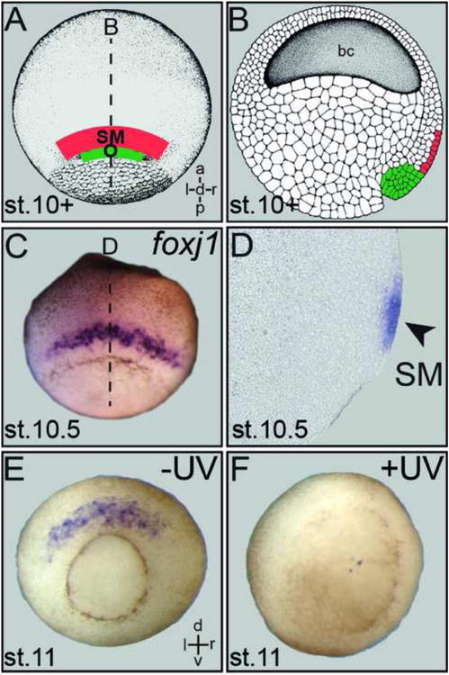
Fig. 2.

Structural and functional relationship of Spemann's organizer and superficial mesoderm. (A) Schematic depiction of superficial mesoderm (SM; red) and organizer (O; green) in whole-mount stage 10+ gastrula embryo shown in dorsal view. (B) Arrangement of organizer and SM in a sagittal section. (C) SM foxj1 expression in a whole-mount gastrula embryo. (D) Sagittal section (plane indicated by dashed line in C) demonstrates foxj1 mRNA in the SM (arrowhead). (E, F) Loss of SM foxj1 expression (E) in UV-ventralized gastrula embryo (F), demonstrating the dependence of SM specification on organizer function.
