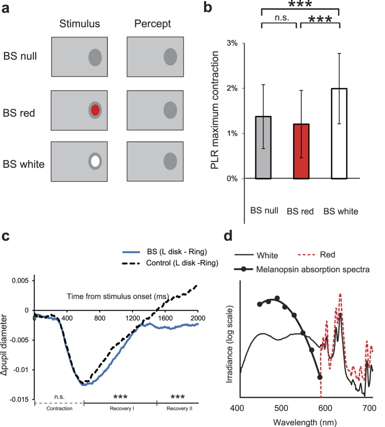
Figure 4. Pupillary light reflex (PLR) in response to light stimulation inside the blind spot (BS) without perceptual filling-in.
(a) Stimulus schematics. Under all conditions, the background luminance increased transiently and yielded a perception of a uniform screen-wide grey, except within the large-disk-sized area around the BS. Under the “BS white” and “BS red” conditions, a white or red small disks was presented inside the BS simultaneously with the background change. (b) PLR size (mean ± 1 s.e.m.) across conditions [N = 4 observers]. *** p < 0.005 (Bonferroni-corrected). (c) Difference in the time course of pupil diameter (Δdiameter) between presentation of the large disk and presentation of the ring at the BS (blue trace) and outside the BS (black dotted trace). ***p < 0.005; n.s. p = 0.70 (ANOVA). (d) Irradiance spectra of the CRT monitor used. The white and red light spectra are plotted against wavelength. The melanopsin absorption spectra are overlaid, adapted from Gamlin et al.22.

