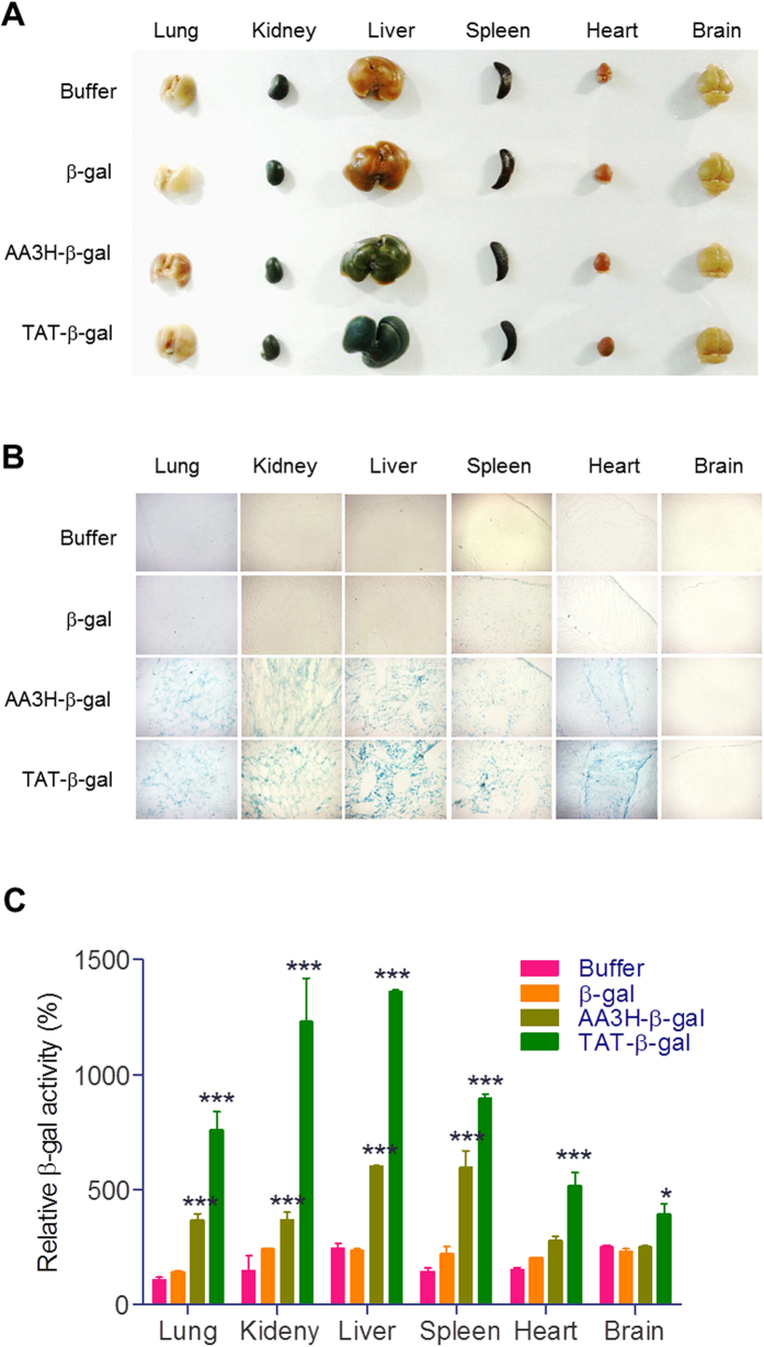
Figure 10. In vivo distribution of β-galactosidase using CPPs.
(A) Harvested organs from mice injected intraperitoneally with the CPP-β-gal conjugates after staining with the X-gal substrate. (B) Sectioned images of each organ shown in (A). (C) Quantitative analysis of β-galactosidse activity in each tissue.

