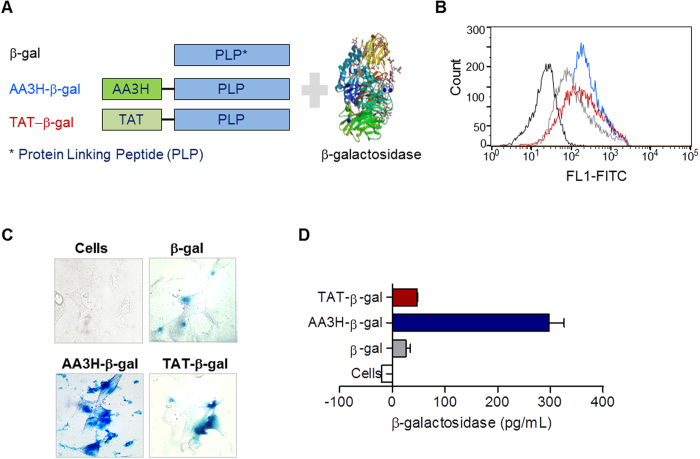
Figure 8. Intracellular delivery of β-galactosidase using CPPs and the activity of the delivered enzyme.
(A) Constructs of CPPs conjugated with the protein linking peptide (PLP) to test the delivery ability of β-galactosidase protein by CPPs into cells. (B) The cells treated with AA3H-PLP (blue trace), TAT-PLP (red trace), or PLP (gray trace) were analyzed on a flow cytometer to estimate intracellular delivery of the peptides. The black trace indicates the untreated cells. (C) Microscopic images of cells treated with the AA3H-β-galactosidase conjugate (bottom left), the TAT-β-galactosidase conjugate (bottom right), or PLP-β-galactosidase conjugate (top right). The image of untreated cells was presented at top left. (D) Quantitative analysis of the activity of intracellularly β-galactosidase by AA3H (blue bar), TAT (red bar), and PLP (gray bar). Activity from the untreated cells was shown with a white bar. Each bar represents the average of three independent experiments.

