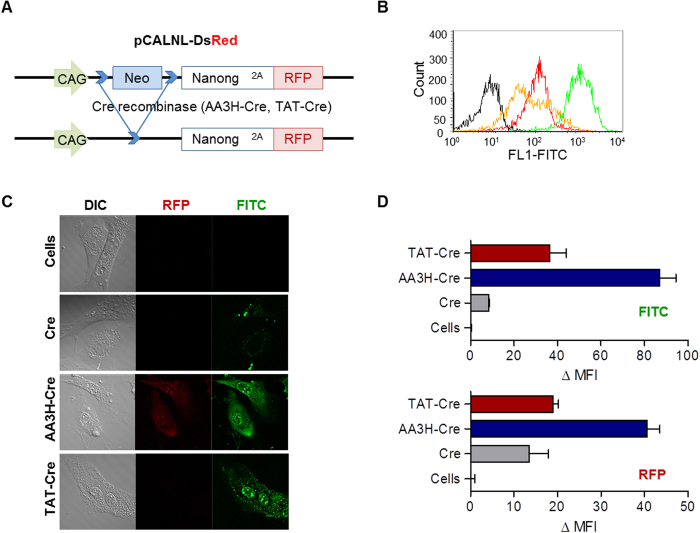
Figure 9. Intracellular delivery of Cre-recombinase using CPPs and the activity of the delivered enzyme.
(A) Constructs of pCALNL-DsRed fibroblast cells to express RFP by cleaved loxP site. Two loxP sites indicated bright blue that holding RFP expression. During recombination by Cre, a stop codon with Neo is removed to lead continuous transcription through RFP gene. (B) Uptake of AA3H-PLP (green trace), TAT-PLP (red trace), and PLP (orange trace) into primary fibroblast cells was analyzed on a flow cytometry. The black trace indicates untreated cells. (C) Confocal microscopic images of cells treated with AA3H-Cre conjugate (third row), the TAT-Cre conjugate (second row), and the PLP-Cre conjugate (second row). Delivery of the conjugates (third column, FITC) and the RFP expression (second column, RFP) due to recombination by the delivered Cre conjugates was monitored. (D) Quantitative analysis of the delivery of the conjugates (top) and the RFP expression by the conjugates (bottom) performed on a flow cytometer. AA3H-Cre: blue bar, TAT-Cre: red bar, PLP-Cre: gray bar, untreated cells: white bar. Each bar represents the average of three independent experiments.

