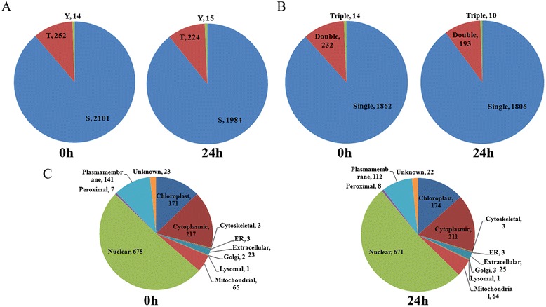
Fig. 2.

The distribution of phosphosite types and subcellular localization of phosphoproteins. a Pie chart showing the distribution of phosphoserine, phosphothreonine and phosphotyreosine. b Pie chart showing the number of phosphopeptides carrying multiple phosphosites. c The subcellular localization distribution of phosphoproteins
