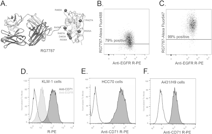
Figure 1. Optimization of flow cytometry method for measuring RIT internalization.
A. Structural model of anti-mesothelin immunotoxin RG7787. Model is hypothetical and previously described in detail13. RG7787 contains a humanized SS1 Fab, shown on the left, linked to a small portion of domain II (processing) and all of domain III (catalytic) of Pseudomonas Exotoxin A through a GGS linker containing a furin cleavage site. Domain III contains 7-point mutations to silence B-cell epitopes. B-C, A431/H9 tumor cell population (anti-EGFR R-PE+) 3 hrs after treatment with 5.85 mg/kg RG7787-Alexa Fluor 488 (B) or RG7787-Alexa Fluor 647 (C). Line is drawn above level at which untreated control tumor cell population has 2-5% positivity (data not shown). D. Histogram showing unstained KLM-1 cells (black, unfilled), stained with anti-EGFR R-PE (gray line, filled), or anti-CD71 R-PE (black line, filled). E. Triple negative breast cancer, HCC70 cells stained with anti-CD71 R-PE. F. Mesothelin transfected epidermoid cancer cell, A431/H9 cells stained with anti-CD71 R-PE.

