
Figure 3.
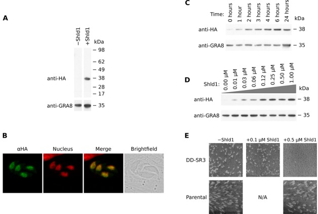
Assays performed on conditional mutants for TgSR3 on a range of Shld1 conditions. (A) Western blot of total protein from purified parasites in the presence and absence of Shld1. Anti-HA was used to detect tagged TgSR3 protein and anti-GRA8 was used as a loading control. Each row is taken from the same blot. (B) Confocal microscopy maximum projections of immunofluorescence assays for mutants in the presence of Shld1. The green channel represents localization of HA-tagged SR protein. The red channel represents the nucleus, as stained by Hoechst. The third pane shows the merge of these two panes, and the brightfield image is last. (C) Western blot of total protein from purified parasites for different durations of incubation with 1 µM Shld1. (D) Western blot of total protein from purified parasites with different concentrations of Shld1 added for 24 h. (E) Plaque assays for conditional mutant and parental parasites. Compared to uninduced transfectants, the plaques of induced transfectants were slightly but significantly reduced in size at 0.5 µM (22% reduction in area; P value = 3.2 × 10⁻⁷). At 0.5 and 1 µM (latter not shown), no plaques were detected. Plaque size of parental parasites were unaffected when 0.5 µM Shld1 was added.