Figure 2.

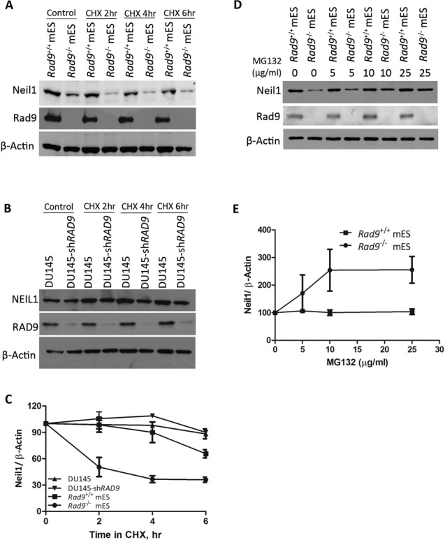
Rad9 controls Neil1 protein stability in mES but not in DU145 cells. (A) Neil1 and Rad9 protein levels were detected by immunoblotting in Rad9+/+ and Rad9−/− mES cells after treating with CHX (50 μg/ml) for indicated time intervals. β-Actin was the loading control. (B) Same as A, but using DU145 cells with or without shRAD9. (C) Average Neil1 protein level relative to β-Actin was calculated by densitometric measurements of bands from three independent experiments (as in A, B). Error bars represent standard deviation. (D) Neil1 and Rad9 abundance was assessed by immunoblotting analyses using Rad9+/+ and Rad9−/− mES cells grown in the presence or absence of proteasomal inhibitor MG132 at concentrations indicated. β-Actin is the loading control. (E) Average Neil1 protein level relative to β-Actin was calculated by densitometric measurements of bands from three independent experiments (as in D).
