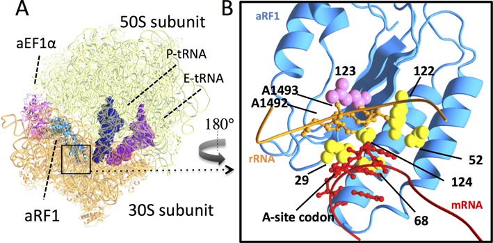
Figure 6.

Putative docking model of the ribosome and eRF1 in a quasi-A/T state. (A) Overall structure. Molecules in the docking model are indicated: aRF1 (light blue), aEF1A (pink), P-site tRNA (dark blue), E-site tRNA (purple), 30S subunits (orange) and 50S subunits (light green) (PDB ID: 3VMF, 2WRO). (B) Detailed view of the decoding center (in the rectangle in (A), opposite view); Domain N of aRF1 (light blue), position-123 (purple) and putative stop codon-binding residues, T29, E52, V68, Y122 and C124, in Saccharomyces cerevisiae numbering (yellow), rRNA (residues 1488–1496, in Escherichia coli numbering) (orange) and mRNA (codon triplets and each of the two bases in the 5′ and 3′ flanking regions) (red). eRF1 residues are shown with S. cerevisiae numbering. Side chains of aRF1 are shown in a sphere model. A1492 and A1493 of rRNA and A-site codons are shown using a ball and stick model. Structural models were rendered by MolFeat version 3.5 (Fiatlux, Tokyo, Japan).
