
Figure 1.
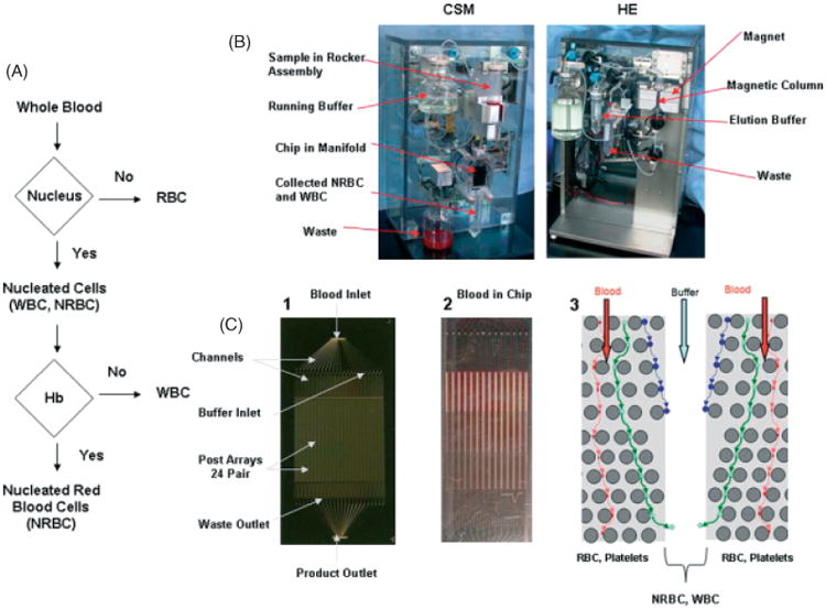
Isolation of NRBCs from whole blood using a microfluidic device. (a) Schematic of flow logic of the microfluidic system. (b) The CSM (left) separates cells based on size through the microfluidic chip into nucleus+ and nucleus− fractions. The blood sample is continually mixed on a rocker, and pumped through the chip using a pneumatic-pressure regulated pump. The HE module (right) collects hemoglobin-containing cells (NRBCs) on a magnetic column that are eluted off the column when the run is complete. (c) (1) A photograph of the microfluidic device etched in silicon, (2) an example of whole blood running through the device, and (3) a schematic of one of 24 sets of buffer inlets and paired array sets on the microfluidic device showing the flow stream of NRBCs and WBCs, deflected by microposts (circles), into the buffer stream. RBCs are small and run into the waste outlets at the bottom of each chip. Colored lines with arrows represent the flow direction of RBCs (red lines), NRBCs (blue lines), and WBCs (green lines)