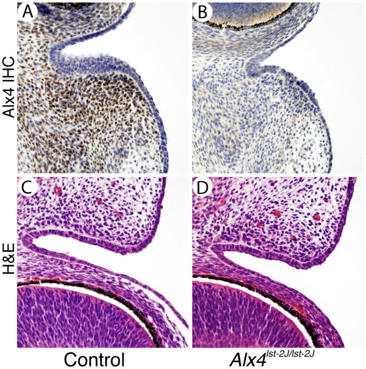
Figure 3.
Expression of Alx4 in the mesenchyme of the developing eyelid. Immunostaining for Alx4 in control eyelids (A) is localized to the eyelid mesenchyme adjacent to the conjuntava. Alx4lst-2J mutants (B) show no specific Alx4 staining in either domain. H&E staining of control and mutant eyelids (C and D) at E14.5 shows normal development of the core eyelid structure at this stage. Scale bar is shown in A is 50 μm and applies to all panels.

