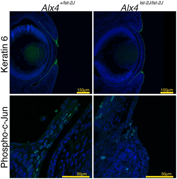
Figure 6.
Expression of markers of eyelid periderm development and activation. Immunofluorescence indicates expression of KRT6 in the clusters of periderm cells at the tips of both WT control (A) or mutant (B) eyelids of E15.5 day embryos. Note the periderm cells in control embryos have expanded and extended partially across the cornea at this stage. (C, D) Expression of Phosph-c-Jun is noted by immunofluorescence in individual cells within the periderm of both mutant and control eyelids.

