
Fig. 4.
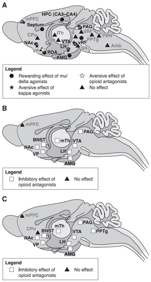
Brain sites where opioid agonists or antagonists modulate drug reinforcement. A: brain regions where injections of opioid agonists have direct positive or negative reinforcing properties. B: brain sites where microinjections of opioid antagonists inhibit the reinforcing effects of opioid drugs given systematically. C: brain areas where local injections of opioid antagonists decrease reinforcing properties of systemic nonopioid drugs (ethanol, cocaine, or nicotine). Reinforcement was assessed using animal models of drug-induced conditioned place preference or self-administration. Brain regions represented on this figure were reported to be sensitive to local opioid injections at least once in the literature. Conflicting results might have been reported in other studies, most often using a different animal model. Noteworthy, most brain areas where opioid manipulations impact on drug reinforcement express all three types of opioid receptors. Amb, nucleus ambiguus; AMG, amygdala; BNST, bed nucleus of the stria terminalis; CPu, caudate putamen; dRF, dorsal reticular formation; HPC, hippocampus; LH, lateral hypothalamus; lTh, lateral thalamus; mPFC, medial prefrontal cortex; mTh, medial thalamus; NAc, nucleus accumbens; PBN, parabrachial nucleus; PAG, periaqueductal gray; POA, preoptic area; PPTg, pedunculopontine nucleus; R, red nucleus; SN, substancia nigra; VP, ventral pallidum; vRF, ventral reticular formation; VTA, ventral tegmental area; vTh, ventral thalamus.