Fig. 5.

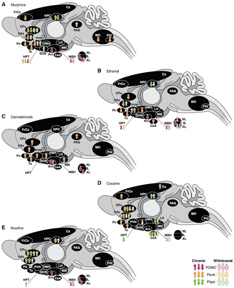
Regulation of opioid peptide genes after chronic exposure to, or withdrawal from, drugs of abuse. Each panel represents brain areas where modifications of endogenous opioid peptide transcript levels were described after chronic exposure to, or withdrawal from, morphine (A), ethanol (B), cannabinoid agonist (C), cocaine (D), and nicotine (E). Chronic drug administration (full arrows) corresponds to repeated injections (constant or escalating doses), pellets implantation, minipump infusions, or self-administration of the drug of abuse. Withdrawal (hatched arrows) was either spontaneous (cessation of injections or removal of pellets) or induced by antagonist injection. Many studies have also reported negative results (no detectable regulation) that are not represented on the figure but discussed in the text. AL, anterior lobe, pituitary; Arc, arcuate nucleus; CeA, central nucleus, amygdala; CPu, caudate putamen; Cx, cortex; FrCx, frontal cortex; HPC, hippocampus; HPT, hypothalamus; IL, intermediate lobe, pituitary; LH, lateral hypothalamus; MBH, medial basal hypothalamus; MM, medial mammillary nucleus; MO, medulla oblongata; NAc, nucleus accumbens; NL, neuronal lobe, pituitary; PAG, periaqueductal gray; PGi, nucleus paragigantocellularis; Pir, piriform cortex; PVN, paraventricular hypothalamus; SON, supraoptic nucleus; Tu, olfactory tubercle; VMH, ventromedial hypothalamus.
