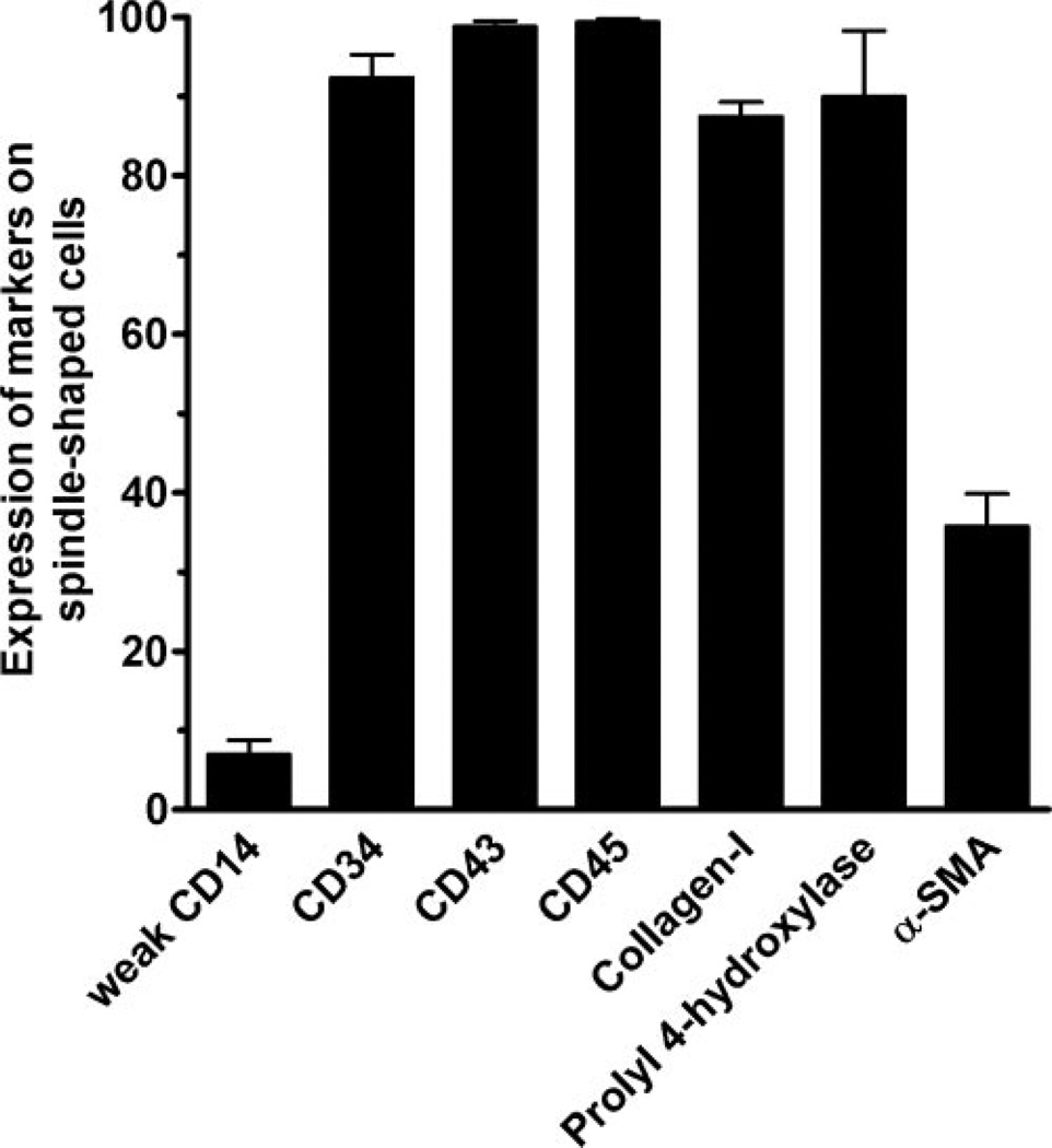
Fig. 2.
Expression of molecules by fibrocytes. PBMC at 2.5 × 10⁵ cells per ml were cultured in eight-well glass slides in serum-free medium for 5 days. Cells were then air-dried, fixed in acetone, and stained with antibodies. Positive staining was identified by brown staining with a blue hematoxylin counterstain. At least 100 elongated cells with oval nuclei were examined from at least 10 randomly selected fields, and the percentage of positive cells is expressed as the mean ± sem (n=3–5 separate donors).

