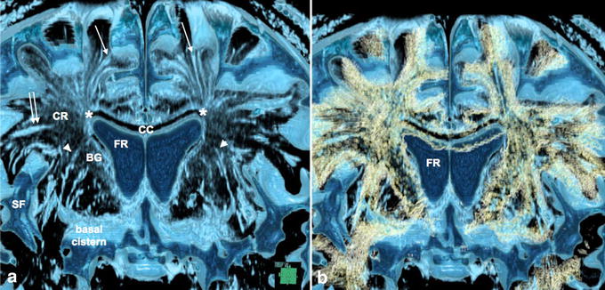
Figure 6.

Virchow–Robin spaces (VRS) in the cerebral hemisphere and association with fiber tracts in a control subject. a Coronal plane volume rendering constructive interference in the steady state (CISS). b Fusion images of CISS and diffusion tensor imaging (DTI) of the cerebral hemisphere. CISS image (a) showed medial VRS (single arrows) in the white matter extending down to the superolateral angle (asterisk) of the lateral ventricle, while lateral ones (double arrow) ended at the lateral end of the corona radiata (CR). VRS in the basal ganglia (arrowheads) ran upward and curved medially to the floor of the lateral ventricle. Fusion image (b) showed VRS in the white matter running parallel to axon tracts (yellow), while VRS in the basal ganglia crossed fiber tracts of the internal capsule. Observation in 69-year-old man, BG basal ganglia, CC corpus callosum, FR frontal horn of the lateral ventricle, SF Sylvian fissure.
