Fig. 7.

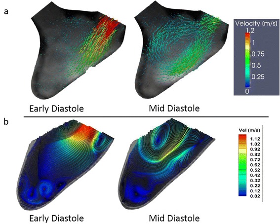
DPIV and PC-CMR measurements on the LV physical model during the early and mid-diastolic phases of the cardiac cycle: (a) PC-CMR velocity vectors (b) DPIV streamlines colored with velocity magnitudes.

DPIV and PC-CMR measurements on the LV physical model during the early and mid-diastolic phases of the cardiac cycle: (a) PC-CMR velocity vectors (b) DPIV streamlines colored with velocity magnitudes.