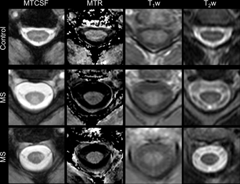
Figure 1.

Comparison of MTCSF, MTR, T1-weighted, and T2-weighted imaging in the cervical spinal cord of a healthy control (top) and two patients with multiple sclerosis (bottom two panels). In the MTCSF images, the deep gray matter “butterfly” is easily distinguished from the surrounding white matter in both the patients and healthy volunteers. Additionally, while obvious on the MTCSF images (arrow), lesions are less conspicuous on the MTR, T1-weighted, and T2-weighted measurements. Figure modified from Zackowski KM, et al, Brain 2009.
