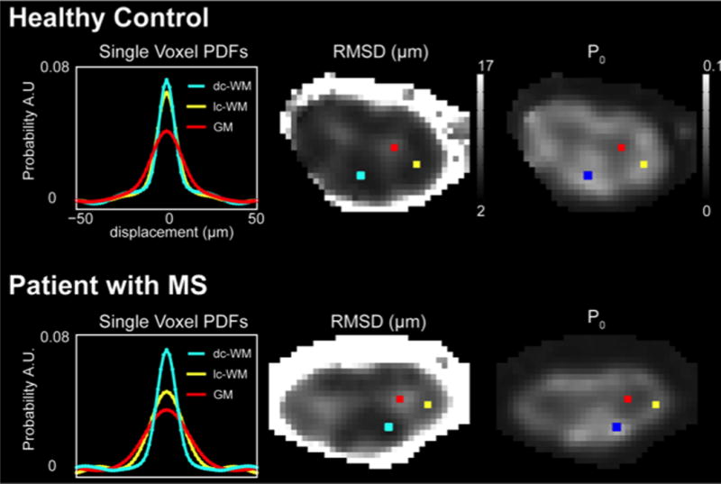
Figure 4.

Demonstration of q-space derived values of root mean square displacement (RMSD) and PDF height (P0) in a healthy volunteer compared to a patient with MS. In the left panel, the q-space derived single voxel PDFs are shown for each of the voxels shown on the images. In normal white matter, it can be seen that a “healthy” PDF is tall and narrow (yellow and blue) while in gray matter (red) the PDF is low and broad. In the MS case with a prevalent lateral column lesion, the PDF for a voxel in the lesion (yellow) approximates the PDF for gray matter (red), while the uninjured column (dorsal column – blue) appears tall and narrow. For the derived RMSD and P0 maps, note that a high RMSD indicates a broader PDF while a high P0 indicates a tall PDF. Figure modified from Farrell JAD, et al, MRM 2008.
