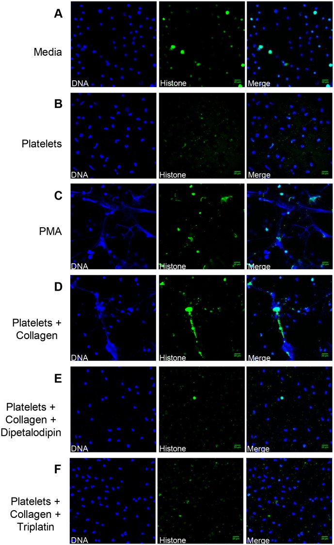
Fig 2. Dipetalodipin and triplatin inhibit platelet-mediated NET formation.
(A-F) Representative images from the immunofluorescence staining for neutrophil activation. NET formation was visualized via confocal microscopy using antibodies against DNA (blue) and citrullinated histones (green), as described in the Materials and methods section. No NET formation was apparent for treatment with (A) culture medium or (B) resting platelets. (C) PMA (5 nM) was used as positive control for the formation of NETs. (D) Treatment with platelets previously activated by collagen (1.3 μg/mL) elicited the formation of NETs. Neutrophil incubation with platelets previously activated by collagen in the presence of (E) dipetalodipin (1 μM) or (F) triplatin (1 μM) did not elicit the formation of NETs. Scale bar: 20 μm.

