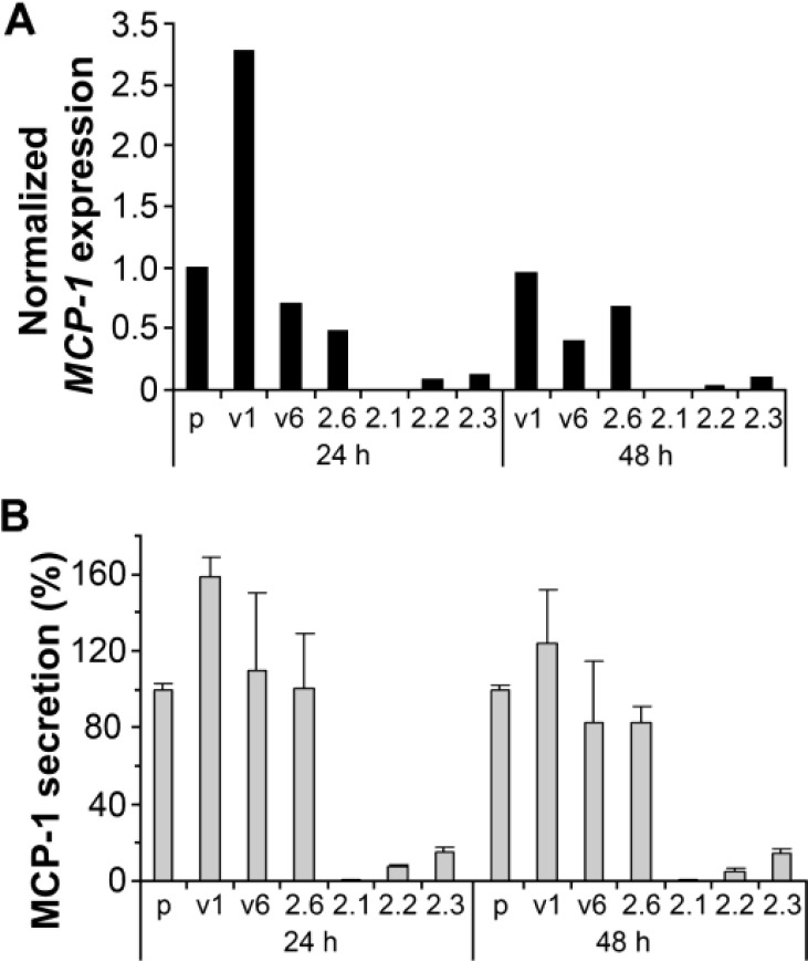
Figure 1. Suppressed expression of CCL2 (MCP-l) in the GBM cell line U251HF expressing transfected PAX6 (2.1, 2.2, and 2.3) as compared with non-transfected (p), vector transfected (vi, v6) and negative PAX6-transfected (2.6) cells.
Cells were grownin DMEM/F12 under normoxic condition with serum deprivation for 24 and 48 hours before getting subjected to the assays. (A) Real-time qRT-PCR measurement of CCL2 expression normalized to ACTB and compared to the level in U25 1HF (p) with serum deprivation for 24 hours, using SYBR-Green I master mix (Roche) and Light Cycler real time PCR instruments following methods described previously 1. (B) ELISA quantification of secreted MCP-1 from the same cells analyzed in A, using MCP-1 ELISA kit from Assay Designs (Michigan). Values include the mean + SD of 3 independent experiments. PAX6 level in each cell line / transfectants by western blot assay was shown in ref. 3.

