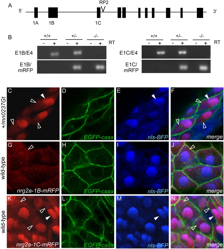
Fig 6. Usage of alternative first exons of nrg2a gene leads to differential cytosolic or nuclear localization of resulting protein isoforms.
(A) A schematic of the nrg2a locus on linkage group 21 (LG21), including alternative first exons 1A, 1B and 1C. The GBT insertion is located in the intron separating exon 1C and exon 2. (B) RT-PCR analyses of 48 hpf sibling embryos from heterozygote (+/mn0237Gt) intercross, revealing that endogenous nrg2a 1B and 1C transcripts are only expressed in heterozygous (+/mn0237Gt) and wild-type siblings (+/+). nrg2a 1B-mRFP and 1C-mRFP fusion transcripts are only expressed in homozygous mutants (mn0237Gt/mn0237Gt) and heterozygous siblings (+/mn0237Gt). (C-N) Confocal images of MFF epidermis at 24 hpf detecting Nrg2a-RFP fusion protein (C, G, K; red), cell membranes (D, H, L; labeled with EGFP (green) after injection of egfp-caax mRNA at 1-cell stage), and cell nuclei (E, I, M; labeled with BFP (blue) after injection of nls-BFP mRNA at 1-cell stage). Panels (F, J, N) show merged images. (C-F) +/mn0237Gt embryo displays Nrg2amn0237Gt-RFP fusion protein both in the cytoplasm (empty arrowheads) and in the nuclei (filled arrowheads). (G-J) Wild-type embryo injected with mRNA encoding exon 1B-version of the Nrg2a-RFP fusion protein; the fusion protein is largely absent from the nucleus, but present in cyptoplasmic compartments. (K-N) Wild-type embryo injected with mRNA encoding exon 1C-version of the Nrg2a-RFP fusion protein; the fusion protein is present in the cytoplasm and the nuclei, similar to the distribution of the transgene-encoded protein (C-F).

