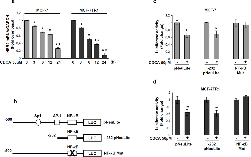
Figure 4.
Effects of CDCA on human HER2 promoter activity. (a) mRNA HER2 content, evaluated by real time RT-PCR, after treatment with vehicle or CDCA 50µM as indicated. Each sample was normalized to its GAPDH mRNA content. *p<0.05 and ** p<0.001 compared to vehicle. (b) Schematic map of the human HER2/neu promoter region constructs used in this study. All of the promoter constructs contain the same 3’ boundary. The 5’ boundaries of the promoter fragments varied from −500 (pNeuLite) to −232 (−232 pNeuLite). A mutated NF-κB binding site is present in NF-κB mut construct. HER2 transcriptional activity in MCF-7 (c) and MCF-7TR1 (d) cells transfected with promoter constructs are shown. After transfection, cells were treated in the presence of vehicle (−) or CDCA 50µM for 6h. The values represent the means ± SD of three different experiments each performed in triplicate. *p<0.05 compared to vehicle.

