Figure 5.

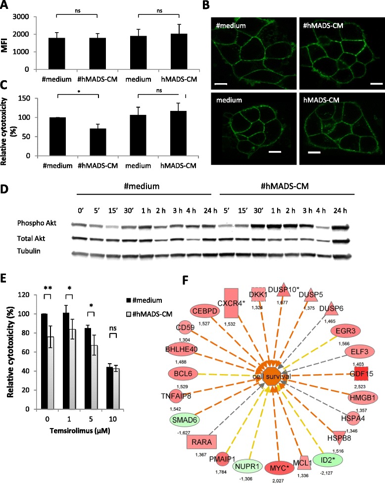
Conditioned media of differentiated human multipotent adipose-derived stem cells increases the resistance of BT-474 cells against ADCC. Human epidermal growth factor receptor 2 (HER2) expression (A) or localization (B) on BT-474 cells after 4-hour incubation with conditioned media of differentiated human multipotent adipose-derived stem cells (#hMADS-CM) or undifferentiated human multipotent adipose-derived stem cells (hMADS-CM) or their control media. Scale bars indicate 10 μm. (C) Antibody-dependent cellular cytotoxicity (ADCC) assays of BT-474 cells preincubated overnight with #hMADS-CM, hMADS-CM or their control media. (D) Kinetic induction of Akt phosphorylation (Phospho Akt) in BT-474 cells exposed to #hMADS-CM or the control medium (#medium) for the indicated times. Quantification of the intensity of the bands is shown. Mean ± SD values of three independent experiments are shown in (A) and (C). Results representative of three independent experiments are shown in (B) and (D). (E) Reversion of #hMADS-induced inhibition of ADCC by temsirolimus. Temsirolimus was added at the beginning of ADCC assays at the indicated concentrations. (F) Genes involved in cell survival in BT-474 cells after exposure to #hMADS-CM. Numbers correspond to fold changes. Vertical rectangle, G protein–coupled receptor; dashed square, growth factor; inverted triangle, kinase; horizontal rectangle, ligand-dependent nuclear receptor; triangle, phosphatase; oval, transcription regulator; trapezoid, transporter; circle, other. Genes in red or green correspond to upregulated or downregulated, respectively. Red lines predict activation; yellow lines indicate inconsistent downstream effect; and gray lines correspond to unpredicted effect. *P < 0.05; **P < 0.01; ns, Not significant.
