
Fig 5. Illustration of signal loss due to motion in in vivo segmented EPI DWI.
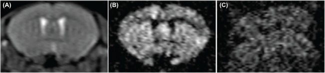
(A) b0 image, (B) and (C) DWI images, all are from the same slice position but were acquired in the presence of minimal (A, B) and excessive motion (C).
Official websites use .gov
A
.gov website belongs to an official
government organization in the United States.
Secure .gov websites use HTTPS
A lock (
) or https:// means you've safely
connected to the .gov website. Share sensitive
information only on official, secure websites.
(A) b0 image, (B) and (C) DWI images, all are from the same slice position but were acquired in the presence of minimal (A, B) and excessive motion (C).