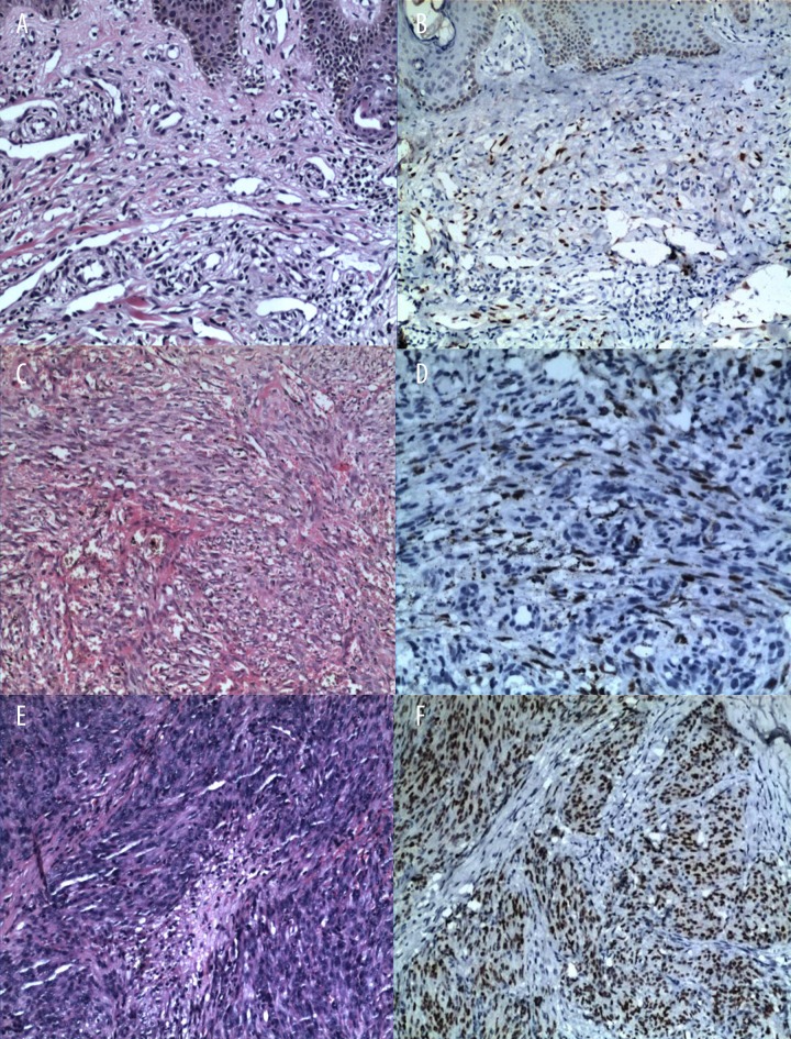
Figure 1.
Representative case of KS in patients of every stage. (A) (Hematoxylin and eosin ×100) showing a macular stage patient. (C) (Hematoxylin and eosin ×200) showing a plaque stage patient. (E) (Hematoxylin and eosin ×100) showing a nodule stage patient. (B–F) The HHV-8 of every stage staining was positive.

