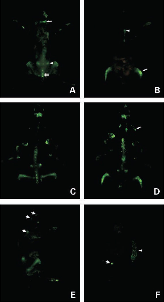
Figure 2.
Whole-body optical images of eGFP-expressing myeloma tumors in skeleton of a live intact mouse. A and B, whole-body, high-resolution fluorescence images of dorsal and ventral views, respectively, of a live myeloma tumor – bearing mouse 4 wks after i.v. inoculation of 106 5TGM1-eGFP cells. A, single arrow, calvaria; arrowhead, iliac crest; double arrows, spine. B, arrowhead, sternum; arrow, hind limb. C and D, corresponding fluorescence images of the exposed skeleton of the same mouse. D, arrow, ribs. E, whole-body fluorescence image of left lateral view of the same mouse. Apart from clearly visible tumors in the femora and iliac crest tumors, fluorescent tumors are also visible in the base of the skull and scapula (arrows). Note the autoflourescent toenails. F, fluorescence image of visceral organs (spleen, brain, liver, kidneys, ovaries, heart and lungs, and adrenal glands) of the same mouse showing tumor infiltration of the spleen (arrowhead) and tumor in one of the ovaries (arrow); all the other organs were tumor-free.

