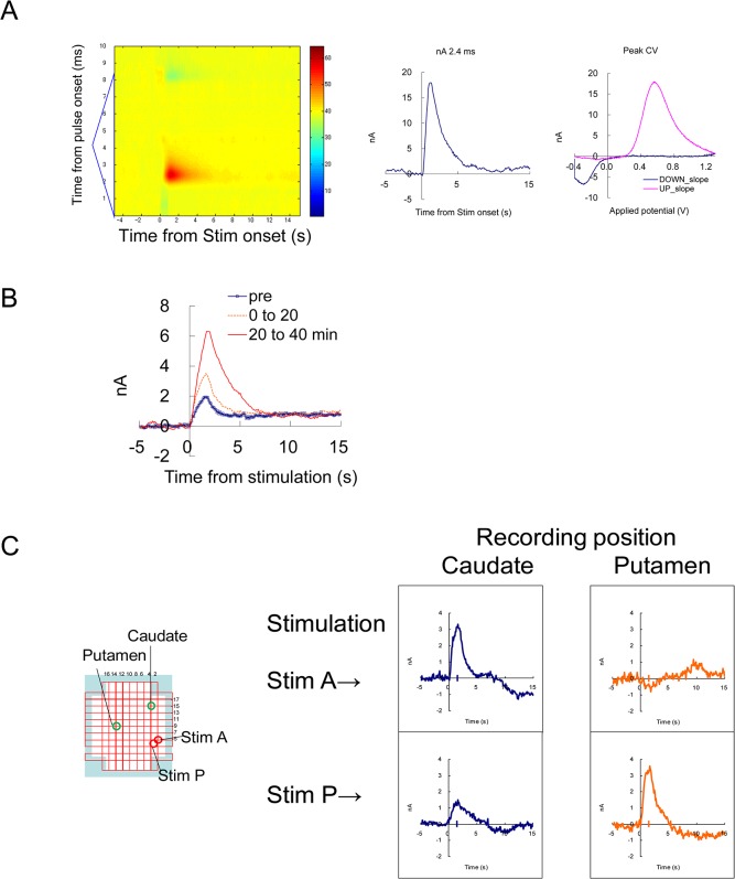
Fig 2. Evoked dopamine release by electrical stimulation.
(A) An example of FSCV recording with MFB stimulation after nomifensine administration. Electrical stimulation of 60Hz, 48 pulses, was given at 5 s from the beginning of the recording. Left: A background-subtracted color plot showing a whole voltage scan from -0.4 to 1.3 V triangular waveform. Middle, Temporal current change at the dopamine peak potential. Right: Cyclic voltammogram from -0.4 to 1.3 V and back to -0.4 V, at the peak of the response. (B) Enhancement by a dopamine uptake inhibitor. MFB stimulation (30 Hz 24pulses) was repeated every 3 min, and an average of 6–7 responses before (pre), up to 20 min (0–20) and 20–40 min after the administration of nomifensine (1 mg/kg s.c.) were indicated. (C) Simultaneous recording from the caudate and putamen using the grid system. Two carbon fibers were placed as in Fig 1E, and two stimulating electrodes were placed in two locations near the MFB. Note that stimulation positions A and P were optimal for inducing responses in the caudate and the putamen, respectively. This result shows simultaneous and independent recording from two locations in the striatum.

