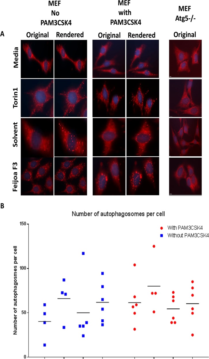
Fig 1. Feijoa F3 activates autophagy in MEF cells.
Cells were treated with feijoa F3 at 10mg/ml, Torin1 at 1μM/ml, with and without 100ng/ml PAM3CSK4 stimulation. A. Shows the LC3B (marker of autophagy) staining. The original images show puncta in cells that represent autophagosomes. The rendered images are created using the Imaris spot function where the spots in the cells are spheres pseudo colored and represent the autophagosomes. The MEF Atg 5-/- showed no puncta B. The puncta were quantified using the spot function in Imaris. Each experimental condition for solvent, media, and feijoa F3 had 3 biological replicates with two technical replicates (giving a maximum of 6 data points) and Torin1 had 2 biological replicates with two technical replicates (giving a maximum of 4 data points). Horizontal line represents median.

