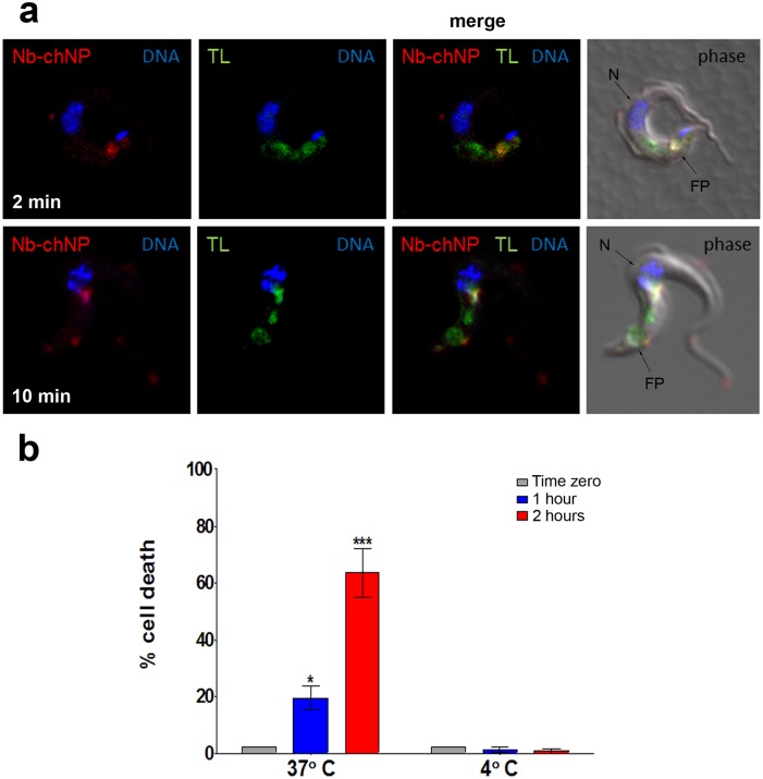
Fig 3. Endocytosis of NbAn33-chNPs.
(a) Bloodstream trypanosomes observed by fluorescence microscopy after incubation with NbAn33-chNPs-Alexa Fluor 594 (red) and tomato lectin-FITC (TL, green) as described in Materials and Methods. Samples were taken after 2 minutes (bottom panel) and 10 minutes (top panel) of incubation. DNA is stained with DAPI (blue). Regions of colocalization appear yellow in merged images. (b) Parasite viability after incubation with NbAn33-pentamidine-chNPs at 37° and 4°C for 2 h. Cell death was estimated by propidium iodide staining and FACS analysis at three time points. Error bars represent the S.D. from three independent experiments. Statistical significance was *, p<0.05; ***, p<0.001.

