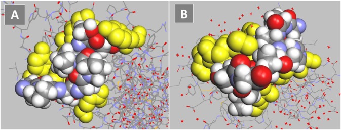
Fig 5. Models of monovalent, mimetic sequence of svH1C (colored space-filled structure) docked in the ligand binding site of receptors (yellow).

(A) Siglec-5 (accession no. 2ZG1), predicted binding energy, -47 kJ/mol. (B) NKG2D (accession no. 1MPU), predicted binding energy, -40 kJ/mol.
