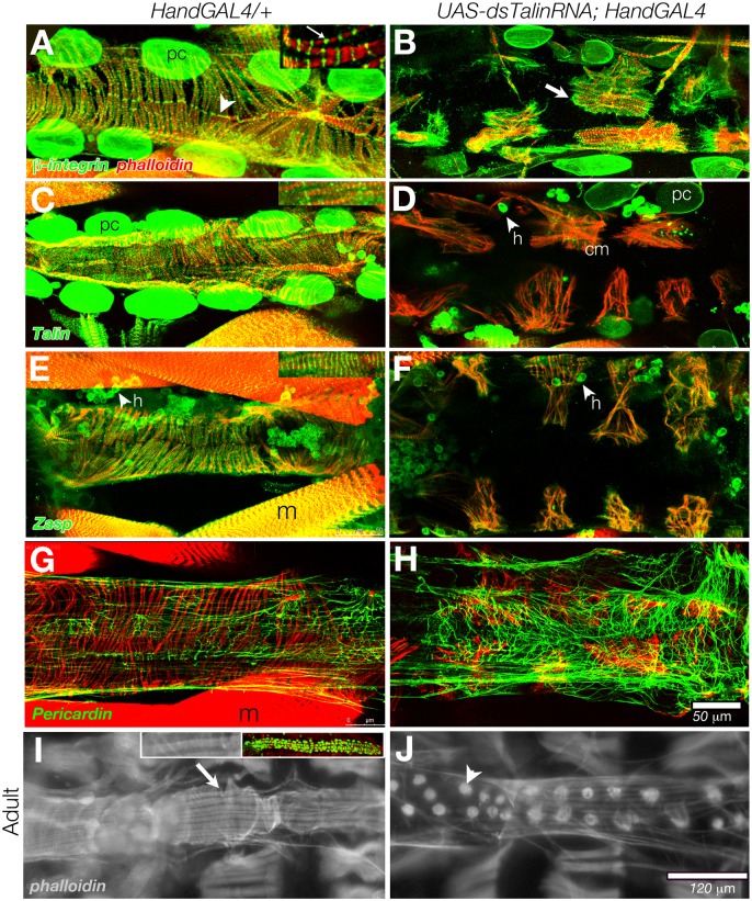
Fig 1. Talin depletion in cardiomyocytes resulted in retraction of the muscle actin network.
The third instar larval hearts lack longitudinal fibres, revealing the transverse fibres (phalloidin, red) and integrin rich cell insertions at the midline. Confocal projections of abdominal segments 5 and 6 of dissected, immunolabled control (Hand-GAL4/+) hearts are shown (arrowhead, β-integrin, green, in A). The contractile heart is flanked by integrin rich pericardial cells (pc). When imaging single myofibrils, Integrin antibody also labeled myocyte Z-lines (arrow in inset, A). When dsTalinRNA expression was generated in UAS-dsTalinRNAi (Hm0516); Hand-Gal4 and examined in third instar larvae, partial myofibril retraction was observed (B). Integrin was concentrated at the cell periphery, beyond the limit of myofibril projection (arrow, B). Talin distribution in wildtype is similar to β-integrin (C), but after chronic depletion in the heart, levels are low in pericardial cells (pc) and cardiomyocytes (cm) but still abundant in hemocytes (h) (D). Zasp, a component of the costamere, aligns to heart and body wall muscle Z-lines, and in hemocytes (E, and inset). This pattern is maintained in myofibrils subsequent to Talin depletion (F). The larval heart ECM contains a latticework of Pericardin rich fibrils (G). After Talin depletion, this latticework becomes more dense in the pericardium (H). The myocytes of the adult control (Hand-Gal4/+) heart are comprised of longitudinal fibres (arrow) overlying the transverse fibres of the cardiomyocytes (I, phalloidin labeling). Relative to the embryonic heart at hatching (inset I, labeled with Hand-GFP), the adult heart has grown 4.7 times in length. dsTalinRNA expression was generated in UAS-dsTalinRNA; HandGAL4 (VDRC 40399) larvae, generating a more severe phenotype, resulting in adult escapers with a collapsed myofibril network, where actin bundles surround each myocyte nucleus (arrowhead, J). Posterior is at right. Scale: 50 μm in A-H; 120 μm in I,and J.

