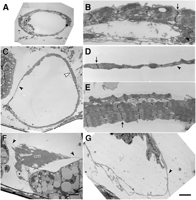
Fig 2. Ultrastructure of Talin depleted larval hearts reveal dilation of the lumen.
The wildtype LIII dorsal aorta (segment A4) was completely encompassed with myofibrils (A, B). Arrows mark Z lines of myofibrils. Extensions of a valve cell line the left and ventral lumen (asterisk, A). After continuous Talin depletion in UAS-dsTalinRNA; Hand-GAL4, the aorta lumen was dilated (C). The aorta wall had zones with myofibril (white arrowhead, C and E) and without (black arrowhead, C and D). Cell processes covered the perimeter of the aorta, and Z-lines are evident (arrowhead, D). Heart cardiomyocytes (cm) retract completely in a severely affected LIII (Segment A6) heart (asterisk, F), exposing naked fibrils of the heart ECM (arrowheads, F, G) The heart in (F) is flanked by fat cells (f). Scale 10 μm (A,C,F) and 1μm (B,D,E,G).

