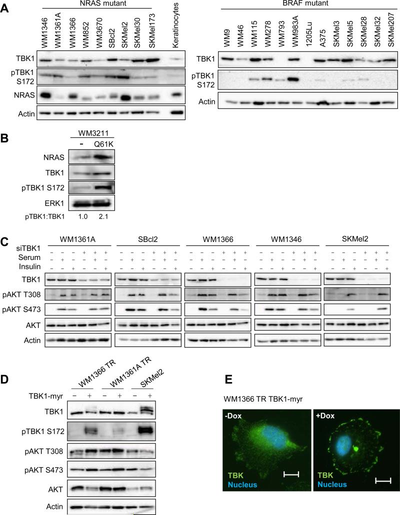
Figure 1. TBK1 is expressed in mutant NRAS cell lines.
(A) Cell lysates from mutant NRAS melanoma cells, a primary human keratinocyte culture, and mutant BRAF melanoma cells were analyzed by Western blot for TBK1, phospho-TBK1, NRAS and actin (loading control). (B) WM3211 melanoma cells wild-type for BRAF and NRAS were non-transduced (−) or transduced with constitutively active NRASQ61K. After selection, cells were lysed and lysates analyzed by Western blot for the proteins indicated. (C) Mutant NRAS melanoma cell lines were transfected with non-targeting or TBK1-targeting siRNA for 72 hours. Cells were serum-starved overnight and treated with serum-free medium, full serum medium, or 1 μM insulin for 20 min. Cells were lysed and lysates analyzed by Western blot analysis. (D) WM1366 and WM136A cells with a tetracycline-inducible system (TR) expressing a TBK1-myristoylated construct (myr) were treated with or without doxycycline for 48 hours; and parental SKMel2 and SKMel2 constitutively expressing TBK1-myr cells were lysed. Lysates were analyzed by Western blot analysis. (E) Immunofluorescence image of WM1366 TR-TBK1-myr with or without doxycycline for 24 hours stained for DAPI (blue) and TBK1 (green). Scale bar = 25 μm.

