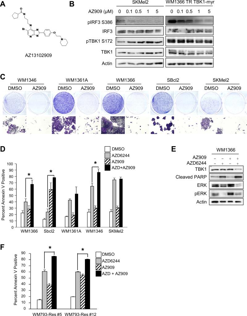
Figure 5. AZ245 in combination with AZD6244 enhances apoptosis in MEK inhibitor-resistant lines in 3D.
(A) Structure of the TBK1 inhibitor AZ13102909 (AZ909). (B) SKMel2 and doxycycline-induced WM1366 TR TBK1-myr cells were treated with increasing concentrations of AZ909 for 24 hours. Cells were lysed and lysates analyzed by Western blotting. (C) Mutant NRAS cells were plated at low density and treated with DMSO or AZ909 (1 μM) for 1 week. Full-sized image, top, and 4× magnification, bottom. (D) Mutant NRAS cells were cultured in 3D collagen in the presence or absence of AZ909 (1 μM) and/or AZD6244 (3.3 μM). After 48 hours, cells were extracted and analyzed for annexin V staining by flow cytometry (n=3; errors bars, S.E.; *, p<0.05). (E) WM1366 cells were placed in 3D collagen and treated as in (D). After 48 hours, cells were lysed and lysates analyzed by Western blotting. (F) WM793-Res #5 and WM793-Res #12 cells grown in 5 μM PLX4720 were cultured in 3D collagen and treated as in (D). After 48 hours, cells were extracted and analyzed for annexin V staining by flow cytometry (n=3; errors bars, S.E.; *, p<0.05).

