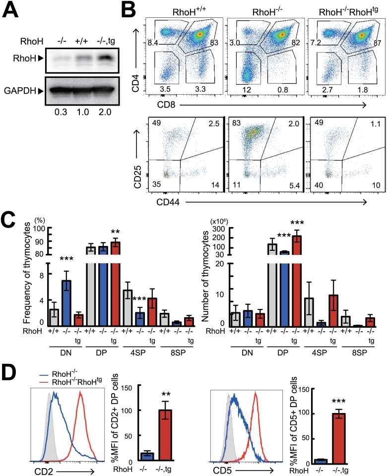
Fig 1. The transgenically expressed HA-tagged RhoH is capable of compensating T cell development in RhoH-/- mice.
(A) Analysis of RhoH protein expression by western blot in RhoH-/-, RhoH-/-RhoHTg, and RhoH+/+ thymocytes. (B, C) Analysis of RhoH-/-, RhoH-/-RhoHTg, and RhoH+/+ thymocytes by flow cytometry. Two parameter plots show CD4 versus CD8 surface staining of thymocytes (upper), and CD25 versus CD44 surface staining on CD4-CD8- (DN) cells (lower). Numbers indicate percentage of cells in the selected area. Bar graphs represent average cell number and frequency of indicated thymocyte subsets calculated from six mice per group. (D) Single parameter histogram plots show CD2 and CD5 staining in DP thymocytes gated on CD4+CD8+ cells (n = 6). Data are shown as mean +SD of more than four mice representative of independent experiments. *P<0.05, **P<0.01, ***P<0.001

