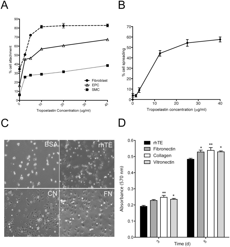
Fig 3. Cell binding to recombinant human tropoelastin.
(A) Relative attachment of human dermal fibrolasts, EPCs and human coronary artery smooth muscle cells (SMC) to increasing concentrations of tropoelastin. (B) The percentage of spread EPCs on increasing concentrations of tropoelastin. (C) Phase contrast microscopy of spreading EPCs on BSA-blocked wells, tropoelastin (rhTE) collagen (CN) fibronectin (FN). Images were taken at 10x magnification. (D) EPC proliferation on days 3 and 5, respectively. Error bars represent S.E.M. of triplicate measurements.

