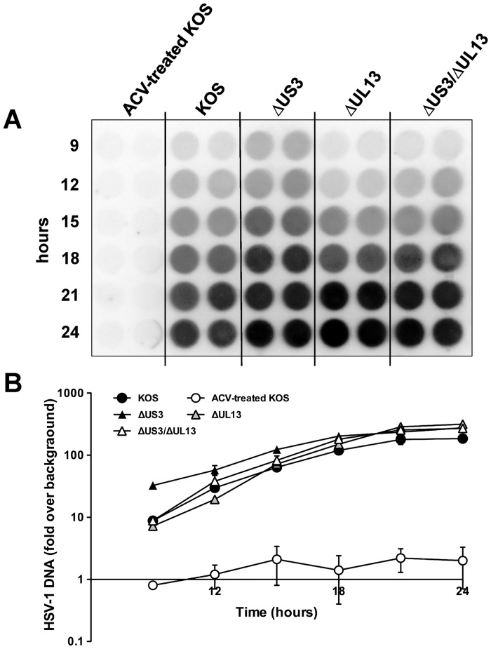
Fig 3. Viral DNA accumulation during the infection with HSV-1 KOS or HSV-1 kinase mutants.
(A) Vero cells were inoculated with wild type HSV-1 KOS or HSV-1 ΔUS3, ΔUL13, and ΔUL13/ΔUS3 mutants at an MOI of 2.5 pfu per cell. At indicated time points, lysates were prepared and blotted onto a nylon membrane. The accumulation of viral DNA was assayed by hybridization to a 32P-labeled US6-specific oligonucleotide probe. ACV-treated KOS served as a negative control. (B) Relative intensity of hybridization signals was quantified by phosphorimager analysis. Results are shown as fold change in hybridization signal intensity relative to the background ± standard deviation (SD) of two duplicate infections.

