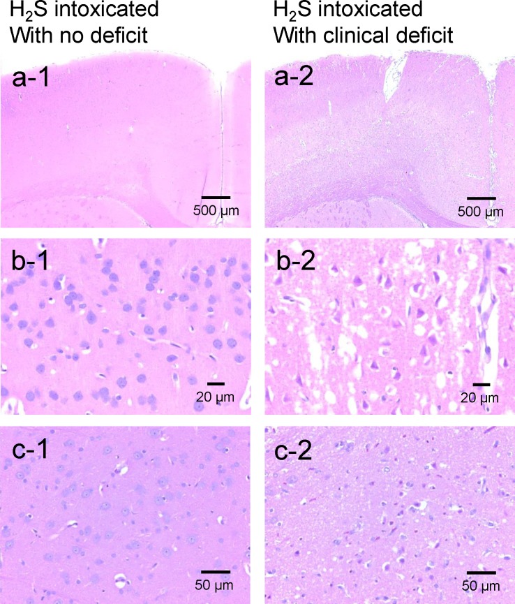
Fig 10. Brain histopathology.
Sections of frontal cortex (panels a and b) and thalamus (c) from one rat that presented a coma but with no neurological deficit (a1, b1, c1) and from rat (#16, Table 1) that was unable to swim after H2S exposure (a2, b2, c2). In contrast to rat with no symptom, the brain of rat #16 showed diffuse and extended neuronal necrosis and neuropil edema affecting the outer frontal cortex (motor agranular cortex) and the cingulate gyrus (anterior limbic area). Neurons are hypereosinophilic with karyolytic or pyknotic nuclei and peri-nuclear edema, Bregma 0.0. 400x. Panel c1 shows normal thalamus at same level and magnification in the intoxicated rat with no deficit. Panel c2: Extensive neuronal necrosis in the lateral posterior nucleus of the thalamus. Bregma -4.8. 400x.

