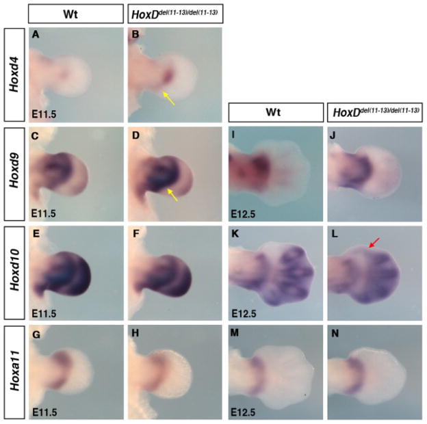
Fig. 1.
Effect of HoxDdel(11–13) deletion on Hox genes expression. A–M: Limb buds hybridized with Hoxd4 (A,B), Hoxd9 (C,D), Hoxd10 (E,F), and Hoxa11 (G,H) at embryonic day (E) 11.5 and Hoxd9(I,J), Hoxd10(K,L), and Hoxa11 (M,N) at E12.5. B,D,J: Yellow arrow points at posteriorly biased expression of Hoxd4 (B) and Hoxd9 (D,J). L: Red arrow point at ectopic second phase expression of Hoxd10 in digit-1 region. Note that the slight difference in the morphology of HoxDdel(11–13)/del(11–13) presumptive autopod at E12.5 prefigures the eventual skeletal phenotype of HoxDdel(11–13) homozygous mice, i.e., shortening of digits and up to six digits per limb.

