
Fig 4. The percentages of apoptotic A549 cells.
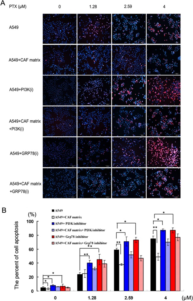
A549 cells were cultured in maintenance medium for 24h and continually cultured in triplicate in the same medium or mixture of maintenance medium and CAF matrix in the presence or absence of the PI3K or GRP78 inhibitor on the 3D chambers for 24h. Subsequently, the cells were stained with Hochest33342 and PI, imaged and the percentages of apoptotic A549 cells were counted. Data are representative images (Fig 4A) (magnification x 200) or expressed as the means ± SD of the percentages of apoptotic cells in individual groups of cells from three separate experiments (Fig 4B). *P<0.05; **P<0.01.