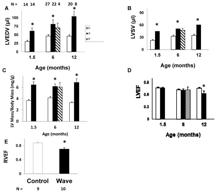
Figure 7. Ventricular structure and function.
A–D: Echocardiography. For Wave mice, data are shown only for mice with moderate or severe aortic regurgitation. E: RVEF, assessed by MRI, at 6 months of age. LV left ventricular, EDV end-diastolic volume, SV stroke volume, EF ejection fraction, RV right ventricular. *p < 0.05 vs. age-matched and treatment-matched Control.

